• Call Now
    + 098370 54507
    0562-2857003, 4000816
s

Dr mathur's ultrasound

Dr mathur's ultrasound colour doppler & x -ray center - Best ultrasound centre in Agra | X- Ray in Agra | best OPG in Agra

We Care About
Your Health

Best Medical Services

24 Hour Service

Dr mathur's ultrasound colour doppler & x -ray center - Best ultrasound centre in Agra | X- Ray in Agra | best OPG in Agra

Read more

Working Hours

  • Mon - web - 10:00am - 8:00pm
  • Thur - 10:00am - 5:00pm
  • Fri - Sat - 10:00am - 8:00pm
  • Sun - 10:00am - 3:00pm

ULTRASOUND AND X-RAY FACILITIES SERVICES

ABDOMINAL ULTRASOUND
OBSTETRICAL ULTRASOUND
TRANSVAGINAL ULTRASOUND
SMALL PARTS ULTRASOUND OF THYROID,EYE,BREAST SCORTUM
OBSTETRICAL DOPPLER
PERIPHERAL VASCULAR
SONOSALPINGOGRAPHY FOR TUBAL PATENCY TEST
INTERNATIONAL ULTRASOUND GUIDED PROCEDURES-ABSCESS DRAINAGE,ASCITIC & PLEURAL FLUID TAP F.N.A.C
service-image

x-ray

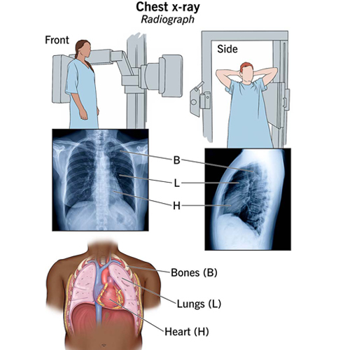
X-ray is a type of imaging technique that uses electromagnetic radiation to see inside the body. It's commonly used to check for broken bones or to diagnose certain conditions. It's like taking a picture of the inside of your body!

Well, an x-ray machine works by producing a controlled beam of x-ray radiation. When the radiation passes through your body, it gets absorbed differently by different tissues. This creates an image that can be captured on a special film or a digital detector. It's pretty fascinating how it allows doctors to see what's happening inside your body without any surgery!

Yes, there are some risks associated with x-rays, but they are generally considered to be very low. The radiation exposure from a single x-ray is quite small, and the benefits of getting the necessary medical information often outweigh the risks. However, it's always important to discuss any concerns with your doctor to ensure that the benefits of the procedure outweigh any potential risks for your specific situation. Safety is always a top priority!

service-image

Ultrasound

Ultrasound technology is a great diagnostic tool for seeing live images of the working structures of the body, especially the structures of joints within the body. Ultrasound imaging (sonography) uses high frequency sound waves, to create a live video feed image of the inside of the body. Ultrasound is the technology, or the “eyes” if you will, for helping doctors get a closer look to make an accurate diagnosis.

Ultrasound is a medical imaging technique that uses high-frequency sound waves to create images of the inside of the body. It's like taking a video of your organs and tissues! It's commonly used to check on the health of a growing fetus during pregnancy, as well as to examine other parts of the body, such as the abdomen or the heart. It's a safe and non-invasive way to get a closer look at what's going on inside.

Well, ultrasound works by using a device called a transducer, which emits high-frequency sound waves. These sound waves travel through the body and bounce back when they encounter different tissues and organs. The transducer then picks up the echoes of the sound waves and converts them into an image that can be seen on a screen. It's pretty amazing how sound waves can give us a visual representation of what's happening inside our bodies!

What Our Patients Says